Xenium: High‑Plex In Situ Spatial Biology at Subcellular Resolution
The Xenium In Situ Platform
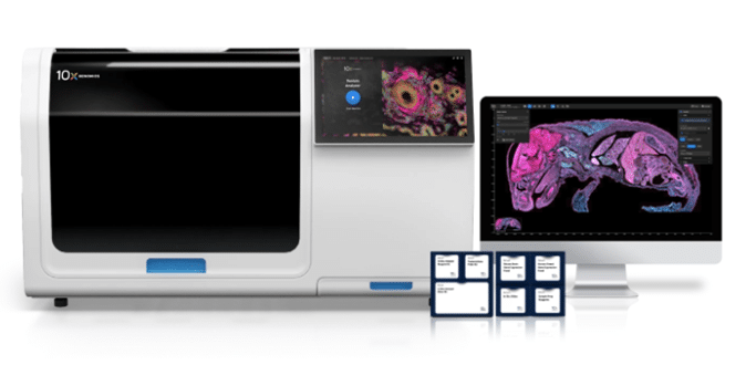
The Xenium platform from 10x Genomics enables subcellular mapping of up to 5,000 genes alongside multiplexed protein expression across intact tissue sections. By leveraging an imaging-based spatial method, Xenium provides subcellular localisation and cell-level insights, allowing researchers to precisely map RNA and protein within individual cells. Designed for high-plex profiling of targeted biology, the platform supports studies in oncology, neuroscience, immunology, and developmental biology. Xenium combines spatial transcriptomics with protein expression analysis in a single workflow, delivering ready-to-analyze data within six days. Data visualisation and analysis are streamlined through the intuitive Xenium Explorer software, making advanced spatial genomics accessible without deep informatics expertise.
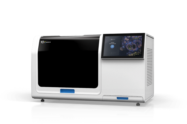
Xenium Analyzer
The Xenium Analyzer is an integrated platform that performs high-resolution imaging, transcript detection, decoding and data analysis in a single automated system. Compatible with fresh frozen and FFPE tissue, it maps hundreds to thousands of genes across large sections with subcellular precision with XY below 30 nm and Z below 100 nm. Flexible throughput options, with 480 genes in under three days or up to 5,000 genes in six, enable scalable spatial transcriptomics and multiomic workflows. Xenium Explorer software provides intuitive visualization and analysis tools to accelerate insights from complex spatial data.
Subcellular detection chemistry
The Xenium platform uses a unique dual hybridization and ligation chemistry that ensures highly specific and sensitive probe binding. This stringent four-factor approach overcomes the off-target issues of single-probe methods and the limitations of probe tiling strategies, delivering reliable subcellular detection across complex tissues.

- High sensitivity comparable to scRNA-seq
- Exceptional specificity ensures low false discovery rate
- Ability to distinguish transcripts with high sequence homology
- Enables interrogation of unique biology, including isoforms
- Reliable detection of short genes and degraded FFPE transcripts
- Tunable detection for highly expressed genes
Xenium slides
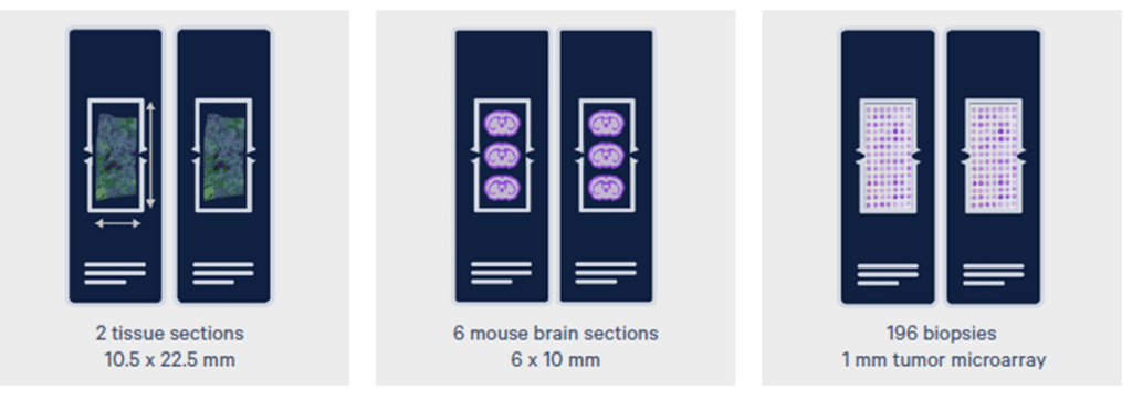
A large analyzable area provides maximum flexibility, enabling profiling of up to 5,000 genes across up to 472 mm² of tissue in six days, with faster analysis possible for panels under 500 genes. Each Xenium slide has an imageable area of 12 mm by 24 mm, with an optimal sample placement area of 10.5 mm by 22.5 mm to avoid covering fiducial markers.
Xenium panels
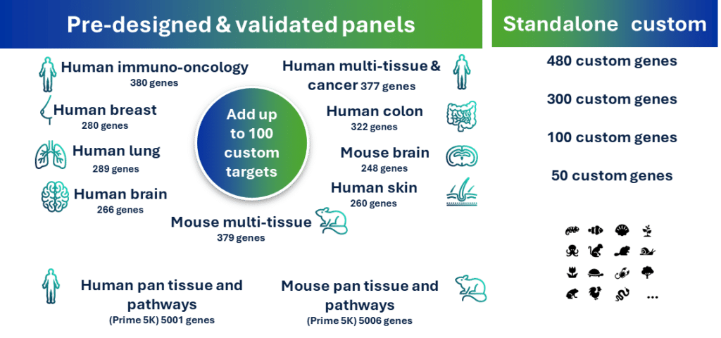
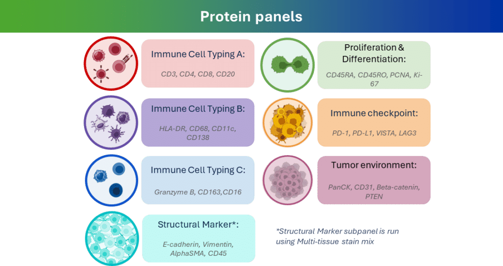
Xenium offers diverse panels to meet any research need. The pre-designed and validated RNA panels have been extensively tested by 10x Genomics on healthy and diseased fresh frozen and FFPE tissues. Xenium also offer custom panel up to 480 genes of your choice. In addition, Xenium Protein subpanels provide ready to use, pre optimised protein marker sets that can be combined with RNA panels for same cell, same section multiomic profiling, revealing both gene and protein expression within the tissue microenvironment in a single workflow.
Streamlined, tissue-agnostic workflow
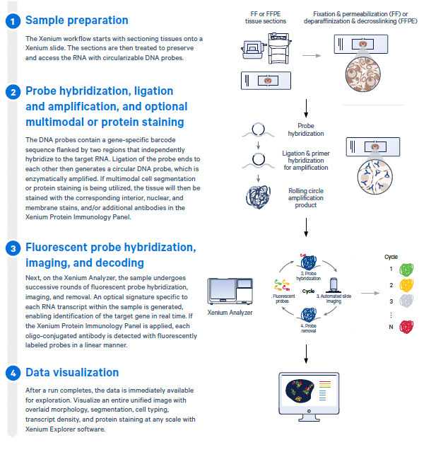
Go from tissue block to instrument-ready samples in just three days with less than six hours of hands-on time. The Xenium workflow enables high-resolution spatial gene expression analysis directly in intact tissue. It begins with tissue sectioning and preparation to preserve RNA integrity, followed by hybridization of gene-specific barcoded DNA probes to target transcripts. Probes are then ligated and amplified, with optional protein staining for multimodal analysis. Prepared samples are imaged on the Xenium Analyzer through successive hybridization rounds, generating fluorescent signatures for each RNA and protein marker. Finally, the integrated data is visualized in Xenium Explorer software, allowing researchers to explore gene expression patterns, cell segmentation, and spatial organisation at single-cell resolution within the tissue microenvironment.
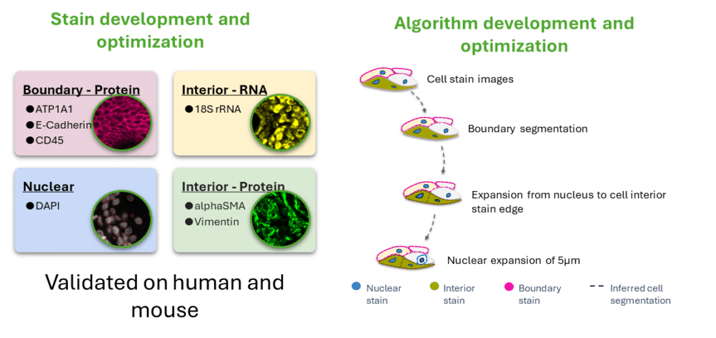
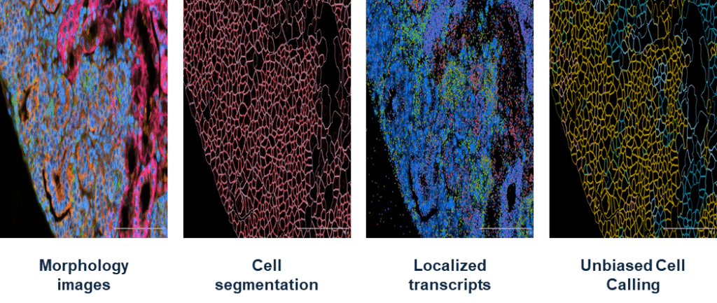
Multimodal morphology-based cell segmentation
Xenium Cell Segmentation provides precise cell segmentation and accurate transcript-to-cell assignment by combining multiple morphological stains with a purpose-built, machine learning algorithm. Optimized staining protocols and advanced algorithms leverage all available image channels to define cell boundaries and capture subcellular features, enabling high-fidelity mapping of RNA and protein expression at single-cell resolution.
Usable data immediately after your run ends
Usable data immediately after your run ends.
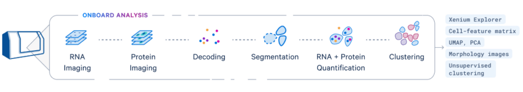
Xenium Onboard Analysis automatically processes data during a run, meaning that interpretable data is ready locally the moment your run is one.
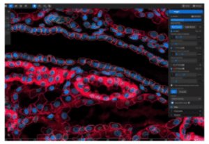
Xenium Explorer, our intuitive visualization software, allows for seamless exploration of your data. Visualize a unified image of your entire tissue section with overlaid morphology, segmentation, cell typing, and transcript density at any scale.
Xenium Ranger analysis pipelines give you the flexibility to further refine your data for your research needs, then continue your analysis journey in Xenium Explorer.
Speak to a Xenium Spatial specialist
On this page
- Xenium: High‑Plex In Situ Spatial Biology at Subcellular Resolution
- The Xenium In Situ Platform
- Xenium Analyzer
- Subcellular detection chemistry
- Streamlined, tissue-agnostic workflow
- Multimodal morphology-based cell segmentation
- Usable data immediately after your run ends
- Speak to a Xenium Spatial specialist